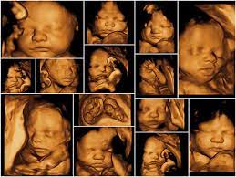
3d - 4d sonography
3D and 4D sonography are advanced ultrasound imaging techniques used in obstetrics and other medical fields to provide three-dimensional (3D) and real-time (4D) views of the fetus or other structures within the body. These techniques offer more detailed and lifelike images compared to traditional 2D ultrasound scans. Here's an overview of 3D and 4D sonography:
-
3D Sonography:
- Three-Dimensional Imaging: 3D sonography creates three-dimensional images of the fetus or the target area within the body.
- Improved Visualization: It provides a clearer and more detailed view of the baby's anatomy, allowing healthcare providers to examine fetal structures, such as the face, limbs, and organs, in greater depth.
- Diagnostic Benefits: 3D sonography can aid in the diagnosis of certain fetal abnormalities or structural anomalies more effectively than 2D ultrasound.
-
4D Sonography:
- Four-Dimensional Imaging: 4D sonography, often referred to as "live 3D ultrasound," provides real-time video images. It adds the element of time to 3D imaging, allowing for a continuous, moving view of the fetus.
- Real-Time Visualization: With 4D sonography, expectant parents can watch the fetus in motion, seeing the baby's facial expressions, movements, and behaviors in real time.
- Emotional Connection: 4D ultrasound offers a more emotionally immersive experience for parents, allowing them to bond with their unborn child by observing their movements and expressions.
-
Uses of 3D and 4D Sonography:
- Prenatal Imaging: Both 3D and 4D sonography are commonly used in prenatal care to assess fetal development, detect anomalies, and monitor the overall health of the fetus.
- Obstetrics: These techniques are used to provide detailed images of the baby's anatomy, measure fetal growth, and assess the position of the placenta.
- Gynecology: In gynecology, 3D and 4D sonography can be used to evaluate the uterus, ovaries, and other reproductive organs.
- Guidance for Medical Procedures: 3D and 4D imaging can assist in guiding certain medical procedures, such as amniocentesis or fetal surgeries.
- Research and Education: These techniques are also valuable tools for medical research and education.
-
Safety Considerations:
- 3D and 4D sonography use the same basic principles as traditional 2D ultrasound, which is considered safe and non-invasive.
- These scans use sound waves to create images and do not involve ionizing radiation, making them generally safe for both the pregnant woman and the fetus.
- However, like all medical procedures, they should be used judiciously and only when medically necessary.
-
Availability and Cost:
- Not all healthcare facilities may offer 3D and 4D sonography. These imaging techniques are often available at specialized ultrasound centers or clinics.
- The cost of 3D and 4D sonography may be higher than that of traditional 2D ultrasound, and it is typically not covered by insurance for purely elective purposes.
While 3D and 4D sonography can provide valuable diagnostic information and enhance the prenatal experience for expectant parents, it's essential to use these techniques under the guidance of a healthcare provider for appropriate medical indications. They should not be solely used for entertainment purposes and should always be conducted by trained ultrasound technicians or sonographers.
Copyright @ Qubeta.